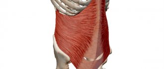
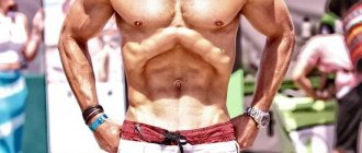
External oblique abdominal muscle[edit | edit code]
External oblique muscle
Home[edit | edit code]
- Eight teeth from the outer surface of the V-XII ribs
Attachment[edit | edit code]
- Linea alba
- External lip of the iliac crest
- Inguinal ligament
- Pubic tubercle
- pubic ridge
Training[edit | edit code]
Read separate article:
Press exercises and training features
Innervation[edit | edit code]
- Intercostal nerves, T5-T12
- Iliohypogastric nerve, T12-L1
- Ilioinguinal nerve, L1
Major injuries and illnesses
All pathologies of the muscular system can be divided into two large blocks: injuries and nerve lesions. Traumatologists and neurologists adhere to strict classifications when making a diagnosis.
Neuromuscular disorders
In neuromuscular diseases, muscle disorders develop due to disruption of the nervous system.
These include:
- Primary and secondary myopathies.
- Congenital myopathies.
- Myotonia.
- Hereditary paroxysmal myoplegia.
- Myasthenia.
Myopathy is a condition in which a muscle undergoes dystrophic changes. In primary myopathies, direct damage to the muscle occurs. It can be caused by inflammation due to a viral or bacterial infection, autoimmune disorders, or mitochondrial failure.
Secondary myopathies manifest themselves as a symptom of pathologies of peripheral nerves. They are congenital, early and late childhood. At first, the disease has no clinical manifestations, so doctors do not immediately diagnose such diseases. As you get older, the disease progresses, gradually leading to muscle atrophy.
Congenital myopathies are distinguished by the fact that at the time of birth, a child’s muscle or group of muscles no longer works. Such diseases do not progress. They cannot be treated, so the child learns to live with his peculiarity. There are no developmental deviations.
Myotonia consists of involuntary muscle contraction. Spasms can be independent or observed in epilepsy. Heredity and stress play a role. Myoplegia has the opposite property: with it, the patient is bothered by sudden attacks of muscle weakness. With myasthenia gravis, the patient has constant muscle weakness. This is usually a congenital condition.
Injuries
Another big block of muscle damage is injuries. They are:
| Closed | |
| Type | Characteristics |
| Bruises | |
| Sprains | The muscle disintegrates, sometimes accompanied by rupture of individual fibers. |
| Breaks | When a muscle is completely torn, its integrity is compromised. |
| Long-term compartment syndrome | a condition that occurs when a muscle is compressed by an external factor (tourniquet or stone) for a long time. Tissue necrosis begins, but due to the obstruction of blood flow, dangerous decomposition products accumulate in the damaged area. When the pressure factor is removed, these substances are released into the circulating blood and enter the vital organs. In the case of the muscles of the abdominal wall, the mere fact of compression is enough to cause death: there is no bone skeleton in this area, so the internal organs are squeezed together with the muscles. The syndrome occurs when buildings collapse, when a person is trapped by massive debris, and rescuers need time to clear a path to him. |
| Open | |
| By application mechanism | Description |
| Wounds | Cut, stabbed, stabbed, torn, bitten, chopped, crushed, bruised, gunshot. |
| Burns | I degree is characterized by damage to the upper layer of skin. Stage II involves damage to the upper layer of skin with the formation of a blister. Grade IIIA is a deep skin lesion; a thin, untouched layer remains at the bottom of the wound. IIIB degree – complete necrosis of the skin down to adipose tissue. IV degree – death of fat, muscle and bone tissue. |
Causes of pathologies
The causes of many neuromuscular diseases are either hereditary characteristics, when such a disease was observed in one of the close relatives, or a violation of embryonic development.
Early childhood myopathies first appear in childhood or preschool age, and late ones occur before puberty. Congenital defects are visible to pediatricians immediately after the baby is born, but usually these affect the muscles of the lower extremities, less often the upper ones. In isolated cases, facial muscles suffer. There is no information about a congenital defect of the abdominal muscles.
External oblique muscle
If myopathy manifests itself in adulthood, then doctors talk about the primary nature of the disease. A virus or bacteria will not immediately infect muscle tissue, because it is not a favorable environment for them. However, inflammation can be transmitted to the muscles if it was in neighboring tissues and was not treated.
Purulent abscesses occur in the subcutaneous fat in the abdominal area and quickly pass through the thin fascia covering the abdominal muscles.
Involuntary contractions or relaxations of muscles in both children and adults have a complex origin. They can be symptoms of serious neurological diseases, or occur due to severe stress experienced by the person.
The abdominal muscles become very stretched during pregnancy. If a woman has an athletic build, then the likelihood of muscle complications after childbirth is greatly reduced. In this case, the abdominal wall is well strengthened, and the muscles are highly developed, so it tolerates internal pressure more easily.
In women who do not exercise their abdominal muscles, the body returns to its normal state more slowly after childbirth. An increase in the waist during this period is normal. If you perform abdominal exercises with moderate load, the abdominal muscles will quickly return to their previous state.
Symptoms
Each pathology has its own symptoms:
Myopathies
The main symptom of myopathies is a decrease in muscle performance. Gradually, it becomes impossible for the patient to use the affected muscles. In the case of the external oblique muscle, it is difficult to rise from a horizontal position, the patient lies on his back with support on his hands, and it is also difficult for him to turn his body in the other direction from the weakened area.
Over time, the muscle begins to atrophy because it is not used. Connective tissue replaces the disappearing muscle. It is dense and inelastic, and to the patient the replacement looks like a trained body.
In this case, contracture occurs - a condition when the muscle cannot stretch. With a contracture of the anterior muscles of the body, it is impossible to bend back or turn.
Myotonia, myoplegia and myasthenia
A myotonic attack is easy for the patient to notice: it is a sharp, strong and sudden contraction of the muscle. If this is not an isolated case, you should consult a doctor.
Attacks of myoplegia also require professional supervision, since relaxation of the muscles involved in the act of breathing can impair a person’s ability to breathe. Such cases are life-threatening. Myasthenia gravis rarely affects the trunk muscles alone. Rather, they will be affected along with the lower limbs and back muscles.
Sprains and tears
The abdominal muscles are painful and limited in movement. Swelling and minor bruising may occur as strains of the external oblique muscle occur during injury. The patient notes that he was engaged in sports and heavy work shortly before the onset of discomfort.
When a muscle ruptures, there is a pathological increase in its function: the area becomes too mobile, since there is no restraining factor. There is a protrusion of the ruptured end, extensive swelling and hemorrhages that extend far beyond the rupture.
Wounds and abdominal bleeding
Wounds that reach muscle tissue are marked by profuse bleeding, which is difficult to stop on your own. In the abdominal area there are no large vessels lying close to the surface of the body, so the patient requires urgent, but not emergency, help. Such injuries do not threaten the life of the victim.
If the wound is caused by a sharp object and cuts the abdominal wall so that through the hole you can get directly into the abdominal cavity, then this condition is dangerous. The integrity of internal organs is damaged and cannot function normally. Such injuries are accompanied by acute pain.
With both wounds and closed injuries, there is a possibility of abdominal bleeding. In the case of small vessels, the patient notes weakness, cold sweat, pale skin and decreased blood pressure.
When larger vessels rupture, the victim experiences severe pain and possible loss of consciousness or shock. With internal bleeding of any scale, a person feels uncomfortable lying down, so he tends to take a sitting position. This is called the “Vanka-Vstanka” symptom.
Diagnostics
Basic methods for diagnosing neuromuscular diseases:
- Blood chemistry. It can be used to determine the level of muscle enzymes: creatine phosphokinase (CPK), myoglobin and aldolase.
CPK is involved in biochemical reactions between a neuron and a muscle fiber. As a result, the nerve impulse is strengthened, and the muscle cell contracts with the necessary force. If the level of CK is elevated in the analysis, this means that the enzyme is released uncontrollably into the blood and is not enough for biochemical transformation. The impulse is not strengthened and the muscle does not contract as it should.
Myoglobin is a protein in muscle tissue that helps transport oxygen. Normally, it is located inside the cell and is not detected in the blood. When myoglobin is released from cells into the blood, the muscles do not receive enough oxygen and cannot function normally.
Aldolase is involved in energy metabolism, its function is the breakdown of carbohydrates. Large concentrations of aldolase are found in muscle fibers, since this tissue requires a lot of energy to work. When aldolase decreases, muscle cells lack “charge.”
- Electrophysiological study. These include electromyography and electroneuromyography. Sensors are attached to the patient to record the force of muscle contractions. At this time, the doctor gives the muscle a load and looks at the instrument readings. This allows you to determine the degree of development of the disease.
- Biopsy. A small piece of the affected muscle is taken from the patient and sent to the laboratory for examination. This method helps to differentiate primary and secondary myopathies.
- Genetic testing. It is carried out in infants when it is impossible to establish verbal contact with the child. Using DNA analysis, geneticists can identify hereditary diseases or chromosomal mutations in a patient.
To diagnose injuries, doctors use:
- Radiography. If a sprain or tear of the abdominal wall muscles is suspected, doctors need to determine whether the ribs have been damaged. A fracture can only be detected on an x-ray or computed tomography scan.
- Ultrasonography. An ultrasound allows the doctor to see abdominal bleeding and assess the condition of internal organs.
- Magnetic resonance imaging. A reliable method for examining soft tissues. Shows a layered image of the scanned area.
Participation in sports[edit | edit code]
As an important rotator of the torso, this muscle takes an active part in athletics exercises - throwing and pushing - in the final phase of movements during which the torso rotates (shot put, hammer throw, javelin, discus). In addition, she performs trunk rotation in kayaking, snowboarding, skateboarding, gymnastics, figure skating, diving, boxing and fencing. When bending the body, it performs both dynamic (fencing and boxing) and static work (maintaining a pose), for example, when shooting from a rifle. With bilateral contraction, this muscle flexes the torso along with the rectus abdominis muscle. Participates in maintaining a straight body position when playing many sports.
| Kind of sport | Movement/hold | Function | Load | Types of abbreviations |
| Shot put, hammer throw, javelin throw, discus throw | Throw phase | Torso rotation | Fast, explosive | Dynamic concentric |
| Kayaking | Turn trunk to support upper limb movements | Torso rotation | Strength endurance | Dynamic concentric |
| Snowboarding, skateboarding | Entering a turn | Torso rotation | Fast, strength endurance | |
| Artistic gymnastics, figure skating, diving | Jumps with a twist or turn | Torso rotation | Fast, explosive | Dynamic concentric |
| Boxing | Participates in attacking strikes, dodging, pendulum movements | Torso rotation | Fast, explosive | |
| volleyball, handball, tennis | Attacking blow, overhead throw, serve, forehand | Torso rotation | Fast, explosive | Dynamic concentric |
| Long jump, high jump, pole jump, triple jump, artistic and sports diving, artistic gymnastics, rowing, football, javelin throwing, contact sports (boxing, judo) | Torso flexion (see “Rectus abdominis”) | Dynamic | ||
| Various sports, such as artistic gymnastics, weightlifting, boxing, fencing, alpine skiing, speed skating, table tennis | Keeping your torso straight | Torso flexion | Strength endurance | Static |
Read also[edit | edit code]
- Muscles - anatomy and functions
- Abdominal muscles
- Core muscles
- Rectus abdominis muscle
- Internal oblique muscle
- Levator testis muscle
- Transverse abdominis muscle
- Quadratus lumborum muscle
- Press exercises and training features
- How to pump up powerful abs
- An effective abdominal training technique
- Abdominal muscle training
- Exercises for the abs
- Abdominal exercises for girls
- Home exercises for the abs
- Cubes on the stomach
- Abs workout for back pain
- Training for terrain
Exercises for the lateral abdominal muscles of men
TRAINING THE LATERAL PRESS MUSCLES FOR MEN
| Exercises | Sets | Repetitions/Time |
| Twisting with lateral transfer of legs | 3-4 | 15-20 |
| Hanging lateral crunches | 3 | 15 |
| Side plank | 1 | from 15 sec. up to 1 min. |
Twisting with lateral transfer of legs
Technique:
- Sit on a flat surface. Press your hands towards your torso. Bend your knees. Slightly tilt your torso back while raising your legs so that the support rests on your pelvis.
- Perform crunches, taking turns moving your legs in the direction opposite to the movement of your torso.
Twisting with lateral transfer of legs
How many:
3-4 sets of 15-20 repetitions.
Read : Effective exercises for the buttocks at home
Hanging lateral crunches
Technique:
- Fix your hands on the bar at shoulder level.
- Pull your legs towards your chest, alternately moving them to the right and left.
Hanging lateral crunches
How many:
3 sets of 15 reps.
Tips: Feet should not touch the floor. Lower them smoothly, tensing your abdominal muscles as much as possible. Beginners can bend their knees slightly to make lifting easier.
Read : How to pump up six-pack abs for girls at home
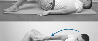
Side plank
Technique:
- Lie on your side, supporting yourself on your forearm. Legs are parallel. Pull the toe towards you.
- Then lift your pelvis off the floor, tensing your muscles so that your body forms a straight line.
- Stay in this position, resting on your elbow and the edge of your foot.
Side plank
How many:
from 15 seconds to 1 minute, depending on preparation.
Tips: Make sure you maintain the correct posture. This exercise is contraindicated for athletes with damaged rotator cuffs, neck muscles, or cervical spine problems.
Read : Abdominal training programs