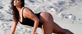
Beautiful, toned buttocks have always been a source of pride for their owners and the envy of those who do not have such roundness. World-famous beauties with “Brazilian butts” shatter the hearts of the stronger sex, or, at least, cause tachycardia in them due to the release of large amounts of adrenaline. Many ladies spend a huge amount of time in the gym, trying to achieve or restore their former elasticity and volume to their buttocks. However, physical exercises, cosmetic procedures and massages are not always able to restore what was lost or acquire something that was not given by nature. There are constitutional features of the figure for which conservative methods are, in principle, not effective. In such situations, plastic surgery comes to the rescue.
METHODS FOR CORRECTING THE FORM OF THE BUTTOCKS
Today, there are several methods in the arsenal of aesthetic surgery to improve the shape of the buttocks, increase their volume and give the necessary elasticity:
- buttock lift with threads (Siluet-Lift, Tissulift and others);
- surgical butt lift;
- installation of implants (buttock replacement);
- lipofilling of the buttocks or, as this method is also called, Brazilian buttock lift.
Regular readers of this site are familiar with some methods of thread lifts from the author’s previous reviews (see the article “Thread lifts”). As for fat grafting of the buttocks, this is a separate large topic, which is also covered on the pages of this site.
In today's review, we will touch upon the anatomy of the gluteal region, ptosis and the mechanisms of its formation, and talk about methods for correcting the shape of the buttocks - surgical buttock lifts. These issues are not sufficiently covered in the domestic literature and therefore, in our opinion, are of great interest to the reader.
How to remove hollows on the buttocks
Modern medicine makes it possible to eliminate even serious defects on the body. How to remove pits on the buttocks? There are several ways to do this.
- In a beauty salon. Modern cosmetology offers a huge list of procedures that can significantly change the condition of the body and skin. The main thing to achieve results is to follow all the cosmetologist’s recommendations and do the procedures regularly until the problem completely disappears. Experts often recommend supplementing the procedures with massage and physiotherapy.
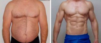
- Power training. This is the most obvious option, which gives results in 100% of cases. Even if your butt dimples do not disappear completely, their shape will be tightened and the fat layer will be reduced. With such buttocks, no one will pay attention to the small pits on them. Active physical activity also helps get rid of cellulite: after several months of exercise, metabolic processes become more active and the skin becomes smoother.
- The surgical method is used in the most extreme cases. There are several options for correcting pits using minor surgery, but all of them do not guarantee the desired result. For example, if you decide to correct your silhouette using lipolifting, the effect may only last for several years.
An experienced doctor will help you choose the option you need. You may have to try several methods to achieve the desired result. Experts recommend starting the search for a solution with simple procedures: physical activity and dietary changes. If these methods do not bring results, you will have to resort to cosmetic and surgical procedures.
ANATOMY OF THE BUTTLE REGION WHEN CORRECTING THE SHAPE OF THE BUTTOCKS
Rice.
1 The structure of the buttocks So, the skeleton of the buttocks is made up of the pelvic bones, the connections between which are provided by dense ligaments, indicated in this figure:
- iliolumbar ligament;
- interosseous sacroiliac ligaments;
- sacro-osseous ligament;
- sacrotuberal (tuberal - ischial tuberosity) ligament;
- superior posterior spine of the iliac crest;
- posterior sacroiliac ligament (Figure 1).
Rice.
2 Gluteus maximus and gluteus medius muscles. The pelvic bones are covered with muscles, over which lies a layer of subcutaneous fat. In particular, this figure shows the large (m.gluteus maximus), and above it the middle (m.gluteus medius) gluteal muscles (Figure 2).
Rice. 3 Superficial muscle layer.
The following figure shows the gluteus maximus (Figure 3, a) and gluteus medius (Figure 3, b) muscles in isolation. Both of these muscles make up the so-called superficial muscle layer. Beneath the superficial muscle layer is a deep layer consisting of several individual muscles (Fig. 4).
Rice. 4 Deep muscle layer.
For greater clarity and consideration of the deep muscles of the gluteal region, it is necessary, relatively speaking, to fold back the gluteus maximus muscle - 1a, 1b in Figure 4. Next: 2 - the middle and, behind it, the gluteus minimus muscles; 3 – pyramidal; 4 – superior twin (m.gemmelus); 5 – internal obturator (m.obturator); 6 - inferior gemellus and 7 - external obturator muscles.
Rice. 5 The location of the branches through which the inertia of the gluteus maximus muscle is carried out.
Innervation of the gluteus maximus muscle is carried out through branches (Fig. 5 - 1,2,3,4) coming from the greater sciatic nerve (6). Number 5 is the pyramidal muscle, from under which the sciatic nerve emerges (6); 7 – coccygeal-tuberal ligament; 8 – ischial tuberosity (tuberal); 9 – greater trochanter (trochanter major) of the femur (Fig. 5).
Rice. 6 Aponeurosis - dense connective tissue membrane
The gluteus maximus and medius muscles are covered with an aponeurosis (a dense connective tissue membrane), over which lies a layer of subcutaneous fat. The uniformity and thickness of this layer determine the smoothness of the transition of the contours of the buttocks (Fig. 6).
There is another important anatomical formation in the gluteal region that is crucial in the formation of ptosis - the subgluteal groove, above which the gluteal fold is formed and held in place. The subgluteal groove is an integral part of the supporting system of the buttocks and is a fusion of the skin and periosteum in the area of the ischial tuberosity (see Fig. 5 No. 8) through dense connective tissue structures. Conventionally, these tight fusions can be called the gluteal ligament, which is part of a single ligamentous supporting system of the buttocks (Fig. 7).
Rice. 7 Multiple dense connective tissue fibers connect the ischial tuberosity and the skin at the very bottom of the buttocks. This structure is responsible for the formation of the gluteal fold and separates the thigh from the gluteal region.
The suspensory ligamentous system of the gluteal region was first partially described by the French surgeon Hypolite Morestin in 1896. He believed that this ligament extends only in the area of the intergluteal fold and perineum due to the intimate fusion of the skin and bone structures of the pelvis through dense connective tissue fibers. In fact, this is a semicircular system that starts from the upper anterior iliac spine, goes through the pubic joint, the perineum, through the ischial tuberosity and ends in the sacrococcygeal joint. Figuratively, this ligamentous fixation system can be represented as a wide, very dense band surrounding the thigh and connecting the pelvic bones and skin. It is surprising that the designers who developed a model of very tight tights with a tightening and supporting effect on the buttocks, probably intuitively, without having a deep knowledge of the anatomy of this area, emphasize and strengthen the ligamentous apparatus described above in their models.
From the above, the following conclusions can be drawn:
- The supporting ligamentous system is not directly related to the muscles of the buttocks and thighs. That is, it cannot be strengthened through physical exercise.
- This very important anatomical formation, which supports and preserves the shape of the buttocks, cannot be destroyed. Rupture of the supporting ligaments (for example, when performing aggressive liposuction in this area) leads to destruction of the ligamentous apparatus and, as a consequence, to ptosis of the buttocks (Fig. 8).
Rice. 8 Ptosis of the buttocks after aggressive liposuction in the area of the upper part of the posterior thigh and gluteal fold.
In the thickness of the subcutaneous fat layer there are many connective tissue transverse bridges (trabeculae), ensuring the compactness of the adipose tissue, low mobility and elasticity of the skin (Fig. 9). Subcutaneous fat is divided into a superficial layer (with more densely and compactly lying fat cells), a middle and deep layer (with large, loose fat cells).
Rice. 9 The structure of the subcutaneous fat layer.
REASONS FOR DEVELOPMENT AND CLASSIFICATION OF PTOSIS OF THE BUTTOCKS
With age, as well as due to decreased muscle tone, sudden weight loss, due to hormonal disorders and changes, connective tissue bridges consisting of collagen and elastin lose their elasticity and stretch. In parallel with this, the volume of fat cells may decrease. These factors together lead to increased mobility and downward displacement of the skin-fat layer under the influence of gravity. In a word – to the occurrence of ptosis (Fig. 10).
Rice. 10 The structure of the subcutaneous fat layer. a – normal, c – ptosis of the buttocks.
The medical term ptosis of the buttocks was introduced by the famous Brazilian plastic surgeon Raul Gonzales. In his opinion, ptosis of the buttocks is excess skin and soft tissue of the gluteal region, which descends below the subgluteal groove.
HOW TO DETERMINE PTOSIS OF THE BUTTOCKS
Ptosis of the buttocks can be congenital (as a feature of the anatomy) and acquired due to sudden weight gain or loss, as well as due to the loss of tissue elasticity and firmness with age and hormonal disorders. The buttocks settle slowly, forming a gluteal fold, the length of which gradually increases from the inner edge of the thigh to the outer. In order to determine the presence of ptosis and its degree, it is necessary to evaluate 2 projections:
The first criterion is the evaluation of the photograph from the front. On the rear projection in front, it is necessary to conditionally draw a line that will divide the posterior surface of the thigh into 2 halves - the posterior midline of the thigh (line M) - and extend it to the buttocks. The second vertical line must be drawn from the ischial tuberosity (tuber) to the sacrum. In the diagrams proposed by R. Gonzales, this is the so-called T-line. Ideally, from the front, the projection of the gluteal fold should not be visible or it should not extend beyond the posterior midline of the thigh. As ptosis of the buttocks progresses, the fold lengthens and in the initial stage extends beyond the midline of the thigh, and then continues to the lateral (outer) part of the thigh.
Rice. 11 Buttocks without ptosis
In this diagram (Fig. 11) there is no ptosis - this is the norm. There is no gluteal fold at all or it only reaches the T line of the ischial tuberosity.
Rice. 12 Minimal preptosis
Minimal preptosis (Fig. 12) - the gluteal fold crosses the T-line of the ischial tuberosity, but does not reach the midline of the thigh - M.
Rice. 13 Moderate preptosis
Moderate preptosis (Figure 13) - the gluteal fold reaches the midline of the thigh, but does not cross it.
Rice. 13 Moderate preptosis
Moderate preptosis (Figure 13) - the gluteal fold reaches the midline of the thigh, but does not cross it.
Rice. 14 Borderline preptosis
Borderline preptosis (Figure 14) - the gluteal fold crosses the midline of the thigh.
Rice. 15 True ptosis of the buttocks
True ptosis of the buttocks (Fig. 15) - the gluteal fold is pronounced and crosses the midline of the thigh at a considerable distance.
The degree of ptosis is determined by a ruler, which is placed in the subgluteal groove and the descent of the gluteal fold is assessed in centimeters (Fig. 16).
Rice. 16 How to determine the degree of ptosis.
- I degree – descent of the gluteal fold 0.5 cm below the subgluteal groove;
- II degree – by 1 cm;
- III degree – 2 cm or more.
The second criterion is profile evaluation. From an aesthetic point of view, it is important to evaluate the femoral-gluteal angle in the profile projection. To do this, it is necessary to connect two conditional lines: the 1st - passes through the point of maximum convexity of the buttocks, and the 2nd - the line of the back of the thigh (Fig. 17).
Rice. 17
As a result, an internal angle is formed, which ideally should not exceed 45°.
Rice.
18 Acceptable (no ptosis of the buttocks) values of the femoral-gluteal angle in the profile projection are no more than 90°.
Fig. 19
As ptosis of the buttocks progresses, the value of this angle will be more than 90°. Accordingly, the more obtuse the angle is formed, the longer the gluteal fold in the front view and the greater the degree of ptosis.
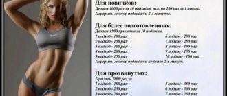
Exercises to correct buttocks at home
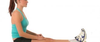
Not everyone has the opportunity to purchase a gym membership, so at home you can also do exercises to tone your buttocks:
- From a sitting position (on a hard surface, back straight), without the help of hands or foreign objects, begin to move forward by tensing the gluteal muscles.
- Lying on your side, gradually raise your leg up, then also slowly down (at least 10 times). Then repeat with the other leg.
- From a lying position on your back, legs bent at the knees, the pelvis slowly rises, stays in this position for 10-15 seconds, then lowers (at least 15 times).
- Standing against the wall, legs extended forward (2 cm from the surface), slowly squat to an angle of 90°, then hold in this position for a minute, then rise.
- From a lying position on your stomach (legs and arms extended), bend your legs, trying to reach your buttocks as much as possible.
METHODS FOR SURGICAL CORRECTION OF THE FORM OF THE BUTTOCKS
To correct congenital or acquired ptosis of the buttocks, a surgical buttock lift technique is used, which has several varieties: upper, lateral, medial (Batterfly) and the so-called DTA lift.
The main problems that can be solved with the help of surgical braces:
- elimination of ptosis (drooping of the soft tissues of the buttocks);
- elimination of tissue sagging;
- elimination of excess skin of the buttocks;
- formation of new shapes and improvement of existing forms.
The method of correcting the shape of the buttocks is selected individually for each patient, depending on the structural features of the buttocks, pursues various goals and is used to tighten different areas of the buttocks, as well as the outer thighs. In the diagrams below, solid lines indicate skin incisions, between which de-epidermization (removal of the surface layer of skin) of skin-fat flaps will be performed. They will further play the role of autoprostheses. The dotted line indicates possible levels of detachment of the skin-fat flap (see below for more details).
Rice. 20 Upper (Pascal or Louran) lift
The upper (according to Pascal or Louran) lift (Fig. 20) aims to tighten the upper outer sections of the buttocks and thighs, and also allows you to create roundness in the upper sections of the buttocks. The technique is effective for significant ptosis of the buttocks and sagging tissue in the “breeches” area.
Rice. 21 Lateral lift and inferior DTA lift
With a lateral lift (Fig. 21) (solid lines of incisions in the flank area), the situation with moderate sagging and flabbiness of soft tissues and skin in the area of the trochanters (large tuberosities of the femur), that is, the outer buttocks and upper-outer thighs, improves. And with the help of the lower DTA-lift (dermo-tuberal anchorage - formation of skin attachment to the ischial tuberosity), indicated by solid lines in Figure 21 in the area of the gluteal fold, you can re-form the subgluteal groove (if necessary in Figure 21), correct excessively elongated buttocks, eliminate asymmetry and mild ptosis, sagging of the lower buttocks.
Rice. 22 Medial buttock lift
Medial (Batterfly) - (Fig. 22) allows you to eliminate mild or moderate ptosis of the buttocks.
The main feature of modern methods of gluteoplasty is that in addition to tissue tightening, it is possible to additionally add volume through autoaugmentation, that is, the creation of autoprostheses from excess of one’s own tissues. This method of operation was first proposed by the famous French plastic surgeons Claude Le Louarn and Jean Louis Pascal. They have shown the benefits of preserving the patient's own tissue to improve the shape of the buttocks and restore lost volume. The previously used methods of traditional dermolipectomy (removal of a skin-fat flap) did not give the desired result, and the buttocks still remained flattened, except that the skin seemed more taut and smooth.
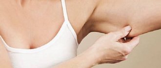
Where do the dimples on the butt come from?
Dimples often appear in women who sit a lot. They can be corrected with anti-cellulite massage. In addition, many exercises have been created that help pump up your butt.
It is worth noting that depressions are a normal part of every “fifth point” of an ordinary person. Everyone has them: men and women, children, old people. To understand how to remove dimples on the butt, it is worth finding out why in some people they are more noticeable, while in others they do not bother them at all.
Beautiful, toned buttocks look sexier on any person, regardless of gender. A saggy or sunken butt can ruin even the slenderest figure. However, if you tighten your muscles through regular exercise, getting rid of the problem will only be a matter of time.
Where do dimples on the butt come from? They appear as a result of fat deposits around certain areas of the buttocks. In thin people they are not noticeable because the subcutaneous cell is evenly distributed under the entire surface of the skin of the buttocks. If the thickness of the layer increases, the depressions become more distinct - there is much less fat in them. The main causes of dimples on the butt:
- hereditary predisposition;
- cellulite;
- injury or surgery.
If you have “bad” heredity, you should start doing special exercises, and the pits will disappear, and the buttocks will acquire ideal shapes. In addition, cellulite can be quickly removed with cosmetic procedures.
OPERATIONS FOR SURGICAL CORRECTION OF THE SHAPE OF THE BUTTHOLES USING THE “BUTTERFLY” METHOD
Rice.
24 The first stage of the operation to correct the shape of the buttocks After making skin incisions along the intended lines and de-epidermization (removal of the surface layer of skin) on the selected “islands,” the ends of the skin-fat flap are separated and unfolded towards each other (Fig. 24, a). The latter are stitched together, forming an “autoprosthesis” (Figure 24, b). This stage of the operation can be reminiscent in its technique of making dumplings, when the corners of the dough are brought together and molded to obtain a completed shape.
The released skin-fat “flap” below the selected “islands” covers the formed “autoprosthesis” (Figure 25, a - shown by an arrow), and the edges of the skin wound are stitched together (Figure 25, b).
Thus, in accordance with the technique of this operation, the skin-fat flap is not simply cut out and removed as redundant, but an “autoprosthesis” is formed from it. Thanks to this surgical technique, in addition to tightening the skin, it is additionally possible to increase the projection, fill the upper part and recreate the lost volume of the buttocks.
TECHNIQUES AND STEPS OF OPERATION FOR CORRECTION OF THE SHAPE OF THE BUTTOCKS
The buttock lift surgery is performed under combined anesthesia (epidural and drug sleep) and takes from 1.5 to 2.5 hours. Before the operation, marking is carried out, the proposed area of the skin-fat “island” is outlined, from which the “autoprosthesis” will be formed (Fig. 26). We will not dwell in detail on the principles of preoperative marking, so as not to overload readers with unnecessary information.
Rice. 27 Preparing the operating team for the start of the operation
Rice. 28 Skin incisions were made according to the intended pattern and the skin-fat “flap” below the “islands” was peeled off
Rice. 29 The stage of de-epidermization of the skin (removal of its surface layer) has been completed.
Rice. 30 Checking the sufficiency of the boundaries of the detachment of the “flap of skin-fat”, which should cover the deep-epidermised “islands”. In this case, the upper and lower edges of the skin wound should freely reach each other without any significant tension.
Rice. 31 The inner (medial) edge of the de-epyremized flap, the “butterfly wing,” is shown, separated from the aponeurosis of the gluteus maximus muscle. The outer edge of the “butterfly wing” is separated in a similar way. This selection of edges must be carried out very carefully and carefully - it is necessary to maintain sufficient vascular nutrition of the “island”, carried out by perforating (penetrating from the depths) arteries.
Rice. 32 The edges of the “island” are released, turned inward and stitched together, as shown in the diagram of Figure 24. This photograph shows that on the left half of the buttocks an “autoprosthesis” has already been formed, resembling an oval in shape. On the right half of the buttocks, the formation of the flap is not yet completed.
Rice. 33 The stage of formation of autoprostheses on 2 sides has been completed.
Rice. 34 The “flap” of the skin-fat flap covers the “autoprosthesis” formed by the surgeon. The photo shows that the upper and lower edges of the wound are compared to each other with temporary adapting sutures.
Rice. 35 The operation is completed, the wounds are sutured in several rows, with an intradermal continuous suture. Active drains (tubes) are installed in the lower part of the buttocks.
Rice. 36 View of the buttocks in profile after completion of the operation. The projection and volume of the buttocks has increased significantly, the gluteal fold has smoothed out, the skin is taut and smooth.
As a rule, after surgery, patients remain in the hospital for one day, and then visit the surgeon for dressing changes on an outpatient basis. It is necessary to wear compression garments for 3-4 weeks, limit physical activity, hot procedures, visits to baths, saunas for 1.5-2 months. Sutures are removed 7-10 days after surgery.
With the lateral and superior lift according to Pascal or Louran, the same techniques are used to form “autoprostheses”. The only difference is the location of the incisions on the skin. Therefore, we did not consider it necessary to go into detail about these butt lift methods.
The technique of the DTA (lower) lift operation has several fundamental differences from previous techniques. And we will be happy to tell you about the nuances of this operation.