Diastasis is an increase in the distance between two sections of the rectus abdominis muscle. The separation occurs at the abdominal suture (Linea Alba and Linea Semilunaris) of the midline connective tissue in the anterior abdomen.
This phenomenon can occur after pregnancy, with rapid weight gain or loss, prolonged and significant physical activity, prolonged cough or regular constipation. The formation of diastasis is promoted by congenital weakness of connective tissue (dysplasia).
Diastasis is often combined with hernias of the anterior abdominal wall of various locations, varicose veins of the lower extremities, flat feet, hemorrhoids, blurred vision and scoliosis.
Strong and prolonged tension of the abdominal muscles leads to an increase in intra-abdominal pressure, and weakness of the linea alba leads to the fact that the muscles, under the influence of the increased intra-abdominal pressure created by them, begin to diverge to the sides. The so-called “vicious circle” closes when muscle tension causes a further increase in diastasis.
During pregnancy, under the influence of the hormone relaxin, the ligaments are stretched and the muscles of the anterior abdominal wall are weakened so that the uterus, and with it the abdomen, can expand with the growth of the fetus. After childbirth, the muscles gradually recover, but the tendons may remain in their previous state and create the appearance of a belly protruding forward.
In addition to a cosmetic defect, diastasis can be dangerous for a woman’s health, since a number of mechanisms that ensure the proper functioning of intra-abdominal structures, abdominal and back muscles are disrupted, and, as a result, pelvic discomfort and prolapse of internal organs may occur .
66% of women with diastasis of the rectus abdominis muscles note the development of urinary incontinence when coughing or sneezing, and discomfort during sexual activity. Diastasis and various pelvic disorders usually go hand in hand.
| 100% of women have some degree of diastasis recti in the third trimester of pregnancy, this means that physiological diastasis cannot be avoided, it is important to monitor the recovery afterwards. |
It is important to note that for many women, rectus discrepancy persists for 6-8 weeks after giving birth, and this discrepancy may remain unchanged for up to a year after giving birth. At the same time, a discrepancy of up to 2 cm is considered a physiological norm.
There are 3 degrees of diastasis depending on the size of the discrepancy of the rectus abdominis muscles:
1st degree - 2-5 cm; 2nd degree - 5-7 cm; Grade 3 - more than 7 cm.
There are three types of diastasis depending on the location of the discrepancy:
- Above the navel;
- Below the navel;
- Mixed type (presence of muscle divergence both above and below the navel).
In 2009, a group of scientists conducted a study - using ultrasound methods, the width of the white line was measured in 150 primiparous women aged 20-45 years.
| It has been found that the average width of the linea alba can vary greatly, from 7 mm at the xiphoid process to 13 mm above the umbilicus and 8 mm below the umbilicus. |
Self-diagnosis of diastasis
If you suspect you have diastasis, we suggest you perform a self-diagnosis using video and record the results. To record, you can use the table in the infographic below or any medium convenient for you.
Diastasis is measured using three parameters:
- Divergence length
Measure how many centimeters the muscles extend up and down from the navel.
- Divergence width
How many centimeters are there between the muscles above the navel and below the navel?
- Finger depth
Evaluate how much your fingers sink when performing the test. During the recovery process, you will learn to engage the transverse abdominis muscle and the depth of immersion of your fingers during the test will become less.
During each test, observe the following aspects:
- Assess whether you experience pain in the pubic symphysis (the joint of the pubic bones).
- Try to feel the boundaries of the muscles along the middle of your abdomen and evaluate whether it is a soft tear or a clear separation of the muscles.
- See if you see a bulge or ridge.
- Listen to the sensations - do you experience discomfort in the pelvic floor (involuntary release of urine, release of gas, air from the vagina).
| If something alarms you while performing these points, you should consult with your doctor to fully assess the condition of the abdominal cavity and/or pelvic organs before starting a cycle of exercises to correct diastasis. |
The video of the self-diagnosis test can be viewed below:
Loads with diastasis
The first thing that comes to the mind of a woman who discovers she has diastasis is to immediately start pumping up her abs. This is a fundamentally wrong decision.
| Classic abdominal exercises not only will not have a positive effect, but can also increase the separation of the rectus abdominis muscles. |
Research suggests that isolated abdominal exercises are not an effective way to combat diastasis .
A 2013 study published in The Journal of Strength and Conditioning Research identifies the following basic isolation and compound exercises that are recommended to be avoided for diastasis:
- lifting the body, legs, both together from a supine position; power crunches lying down, “bicycle”, “scissors”;
- yoga asanas that create strong pressure on the midline of the abdomen, such as Mayurasana and the like;
- power poses that significantly increase intra-abdominal pressure - power balances on the arms, swings and kicks, jumping, push-ups;
- poses that stretch or expand the abdominal wall - deflections, variations of bridges, including on a fitball or bench for deflections , pull-ups;
- straight planks;
- long stands on hands/elbows and knees/feet, such as push-ups.
| In everyday activities, you should avoid stress that leads to an increase in intra-abdominal pressure. Therefore, you have to think about redistributing household chores for a while. |
Please note a few important points:
- Wear a postpartum wrap when carrying your baby, especially if it lasts for a long time
- Do not carry weights weighing more than 5-6 kg . When lifting objects, bend your elbows rather than holding them at arm's length:
| Right | Wrong |
- Lie down and get out of bed on your side , as during pregnancy. Sleep on your back or side.
- If you need to get up from a chair , pull in your stomach and stand up mainly using the strength of your legs.
- Maintain correct posture: do not arch in the lower back, but do not slouch either.
Babywearing can also make diastasis worse. It doesn't matter whether you carry your baby in a sling, baby carrier, baby carrier, or in your arms, as long as you're violating your posture. It is poor posture in most women that causes pain in the lower back, thoracic spine, and pubic symphysis and provokes an increase in the degree of diastasis .
The main mistake many women make is rounding their back, with their shoulders moving forward and the pelvis also moving forward - this compensates for the weight of the child by shifting the center of gravity, but significantly harms the health of the mother. In addition, the very weight of the child leads to an increase in intra-abdominal pressure, provoking an even greater divergence of the rectus abdominis muscles.
If you experience discomfort or pain while carrying a child, or you have grade 2-3 diastasis, you should stop using a sling during rehabilitation.
| If you are just planning a pregnancy, to prevent the development of significant diastasis, you should pay attention to strengthening the abdominal muscles, pelvic floor muscles, deep back muscles and diaphragm. |
During pregnancy, this will help support the growing belly, prevent pain in the lumbar spine and prevent prolapse of the pelvic organs. It has also been noted that with good development of deep stabilizer muscles, recovery after childbirth is much faster.
What is diastasis from an anatomical point of view?
Diastasis is an increase in the distance between paired rectus muscles, namely their moving away from each other due to stretching of the linea alba. Let's look at this state in more detail:
- Each of the muscles of the abdominal wall is covered with a sheath of connective tissue - an aponeurosis.
- The plexus of fibers of the aponeuroses of all muscles forms the linea alba, which is located from top to bottom in the area between the xiphoid process and the pubic symphysis.
In other words, the distance between these paired muscles on the abdomen with diastasis is greater than is normally necessary, as a result of which difficulties arise in strengthening the abdominal press. This occurs due to overstretching of the white line of the abdomen, which becomes thinner and does not seem to return to its original position.
How to quickly and accurately determine whether there is diastasis?
Externally, this condition manifests itself as a protrusion in the form of a roller or a dip when the abdominal muscles are tense in the midline.
It is quite easy to independently determine the presence of separation of the rectus abdominal muscles. To do this, you need to lie on the floor and lie on your back, slightly bend your knees, raising your head. In this posture of the body, when the rectus abdominis muscles are tense, diastasis is diagnosed by assessing the distance between them with a finger.
Normal condition: at the level of the lower ribs - 15 mm, at the level of 3 cm above the navel - 22mm, at the level of 2 cm below the navel - 16mm. It is also important to evaluate not only the width of the discrepancy, but also the depth and density of the white line. Connective tissue density is more important for health. For beauty - density and distance.
If you suspect that the muscles have separated, you should seek help from a surgeon, who will conduct an examination and prescribe an ultrasound examination to determine the width of the white line, exclude an umbilical hernia, etc.
Where does diastasis come from?
Diastasis of paired muscle fibers of the abdominal wall can occur in the presence of one or more of the listed reasons. It can develop in newborns, more often premature babies, and also in men. The cause may also be a genetic feature of connective tissue. Let's consider the reasons for the discrepancy of the rectus abdominis muscles in women.
Pregnancy
The most common cause of separation of the rectus muscles on the abdomen is pregnancy, in which, due to the growing uterus, intra-abdominal pressure increases and, accordingly, the load on the anterior abdominal wall.
Starting from the second trimester after about 14 weeks, when the fundus of the uterus is located approximately 10 cm above the pubis, the process of divergence of the rectus abdominis muscles begins. In the third trimester of pregnancy, increased extensibility of the linea alba is observed in at least 60-70% of pregnant women, while diastasis can increase in size until the birth itself. Predisposing factors to divergence of the rectus abdominis muscles during pregnancy are:
- woman's age over 45 years;
- overweight;
- pregnancy with a large fetus or multiple pregnancy;
- rapid weight loss;
- high physical activity in the first months after childbirth.
Increase in the hormone relaxin
During pregnancy, a woman's hormonal background changes, resulting in a decrease in the strength of the collagen fibers of the connective tissue.
This occurs due to an increase in the synthesis of the hormone relaxin, which is produced by the body to relax the muscles in the pelvic area and helps to expand the cervical canal to prepare the birth canal for the birth of a child. Relaxin affects the connective tissue fibers between the pubic bones, which is often accompanied by their divergence and is called symphysiopathy. That is why we can safely say that during pregnancy the development of diastasis is promoted not only by an increase in intra-abdominal pressure, but also by physiologically determined changes in hormonal levels. Today Live training with Tatyana Metelskaya Instagram
Full body fat burning workout 19:00 Free workout
View schedule
C-section
Almost any abdominal surgery in the area of the anterior abdominal wall in one way or another affects the white line of the abdomen.
During a caesarean section, when spontaneous childbirth is impossible, an incision is made above the pubis perpendicular to the midline, disrupting the integrity of the aponeurosis and making it more susceptible to stretching. In the postoperative period, the size of the uterus gradually returns to its original size, the abdomen decreases in size, but the damaged linea alba is difficult to restore. Stages of diastasis depending on the divergence of the rectus abdominis muscles:
- Stage 1: distance of 2.5-5 cm, symptoms are mild, diastasis is faintly noticeable in appearance.
- Stage 2: distance by 5-8 cm, the muscles in the abdominal area are weakened, the skin is sagging.
- Stage 3: a distance of 8.1 cm or more, during which prolapse of internal organs and disruption of their functioning is possible.
If the pathology is not eliminated in a timely manner, as it progresses, the following may occur:
- problems with posture;
- fast fatiguability;
- gastrointestinal disorders with the appearance of heartburn, belching, flatulence;
- minor pain in the pit of the stomach.
After childbirth, at least two months must pass for the muscle aponeurosis to recover. If diastasis persists a year after the birth of the baby, independent restoration of the anterior abdominal wall to its original state is unlikely.
What exercises are contraindicated for diastasis?
All exercises that provoke excessive stress on the rectus abdominis muscles, as well as exercises that provoke excessive pressure on the anterior abdominal wall, are contraindicated. It is not recommended to do:
- classic abdominal exercises: crunches with shoulder or leg lifts;
- push ups;
- classic plank;
- deep squats and lunges;
- strong deflections and stretching of the anterior abdominal wall;
- abdominal breathing techniques;
- training with heavy lifting, etc.
Surgical correction
| Abdominoplasty is a surgical method for eliminating diastasis. For this purpose, tension plasty with local tissue or non-tension plasty with a synthetic mesh endoprosthesis are used. |
Current scientific evidence and clinical experience indicate that biomechanical and physiological causes, pregnancy and childbirth can affect the fascia of the abdominal support system, leading to diastasis. But not all women with diastasis recti require surgery to restore full function.
The current clinical hypothesis is that at least one year must pass after delivery before a decision can be made about the need for surgical correction of the dystasis.
| Finally, surgical intervention can only be indicated if a multi-tasking system for correcting dystasis, including nutrition, exercise and an active lifestyle, has proven ineffective for recovery. |
Causes
Most often, the problem worries women; diastasis of the rectus abdominis muscles after childbirth appears in almost every fourth woman in labor. Miniature mothers of two or more children who have a delicate constitution are especially susceptible to excessive stretching of the aponeurosis of the rectus muscles. Women who were involved in sports before pregnancy and have a thick build are least at risk. Diastasis of the rectus abdominis muscles in men often appears due to an incorrectly selected set of strength exercises, and in young athletes due to coaching errors. The constitutional characteristics of children can provoke the onset of the disease in the absence of an individual approach to the choice of exercises. The so-called dysplastic children have a congenital weakness of the connective tissue, which also makes up the bridge (aponeurosis) between the muscles. Diastasis of the rectus abdominis muscles in a child: with existing weakness of the connective tissue, with constant loads leading to increased intra-abdominal pressure, an abnormal divergence of the superficial rectus abdominis muscles is formed. Young dysplastic athletes can be identified by a number of characteristics:
- detect MAPSS syndrome - minor cardiac anomalies;
- valgus shape of the lower extremities (X-shaped legs);
- flat feet with valgus deformity (most of the load falls on the inner edge of the foot);
- frequent ankle subluxations.
In pregnant women, a temporary state of thinning, “softening” of tissue along the midline of the abdomen results from:
- increased intra-abdominal pressure due to a growing uterus;
- Hormonal changes contribute, which contribute to greater compliance of the ligamentous apparatus of the pelvic bones (for the safe passage of the fetus through the birth canal).
Repeated births with diastasis of the rectus abdominis muscles lead to an even greater intermuscular defect. Often, women, in an attempt to regain their previous shape, resort to rigorous strength training, but an incorrectly selected set of exercises for diastasis of the rectus abdominis muscles after childbirth leads to great disappointment - the discrepancy of the muscles can increase due to increased intra-abdominal pressure during exercise. And the annoying bulge of the abdomen will not disappear, because it is impossible to train the aponeurosis (tissue between the muscles) - there are no muscle fibers there and it will not decrease after physical exercise. exercises.
Exercises to correct diastasis
It is important to note that the approach to correcting diastasis through exercise should be gradual and regular . Our system involves gradually engaging the deep core muscles to ensure an even load. As a result, this approach leads to the closure or significant reduction of diastasis.
| You can perform a set of exercises to correct diastasis in grades 1 and 2, in the absence of hernias and no earlier than 6 months after birth by cesarean section and 1 month after natural birth (start from the first stage of exercises). |
If you have grade 3 diastasis, you should stop at the first stage of the program until it reaches at least grade 2.
The #sekta diastasis rehabilitation program is conditionally divided into two stages:
- the first stage is the formation of a physical activity regime;
- the second stage is the transition to more intense exercises.
Work on correcting diastasis begins with correcting or strengthening posture, walking, joint exercises, normalizing diet, breathing exercises, strengthening the transverse abdominal muscle and pelvic floor muscles.
Video: how to check diastasis
Wondering if you have diastasis? Watch this video to test yourself and determine if you have an abdominal muscle discrepancy!
Below are my belly photos from 38 weeks pregnant to 10 days postpartum. While every woman's postpartum body changes/heals/bounces back differently, I'm simply documenting my journey in hopes that it will help other moms cope with diastasis.
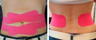
As you can see, my belly has shrunk dramatically in a short period of time. I owe this to the postpartum corset, which I wore non-stop (all day and night), as well as postpartum basic exercises. It's been two months since my second child was born and I'm still wearing a corset. While I love the benefits of this bandage, simply wearing it alone will not stop your diastasis from appearing. Stabilizing your core muscles and strengthening your transverse abdominal muscles are also absolutely essential to getting your body back to normal.
Hold and tighten the elastic, wrap around your stomach and secure
Naturally, the degree of tightening of the corset should vary depending on the time elapsed after childbirth. And, as a rule, they are specially made so that they fit both on the first day after birth and after 3 months.
Would I recommend a corset for pregnant women and new mothers? Absolutely. Not only do I know for sure that it helped shrink my belly, but it also provides very good support for my lost core muscles. In addition to diastasis, I had an umbilical hernia (also common during pregnancy), which bothered me during the last few months of pregnancy and the first few weeks after the baby was born. Although the hernia is still in my body (and will remain), it has not caused me pain yet, thank God. I believe the bandage and training in a quality and effective set of exercises, which is pictured below, helped me. Here are the RIGHT exercises to do to eradicate diastasis.
Correct posture
One of the most important, although seemingly insignificant exercises, is developing the skill of correct posture.
Over the course of some time, you will need to constantly monitor your body position: shoulders back, level above the pelvis, stomach moderately tucked, hips level, feet shoulder-width apart, toes spread for stability.
At first it will be unusual, but over time you will form the habit of standing and moving without disturbing your vertical position. Try not to tense your facial muscles, so as not to cause tension in your body. When moving your ribs and shoulders, you should not feel any discomfort in your stomach or hold your breath.
Make sure that your tailbone doesn't move forward and that your weight is evenly distributed without shifting to one hip - all these pelvic tilts indicate that the transverse abdominis muscle is not working. To engage the transverse muscle, pull your belly toward your lower back, concentrating on the area below your belly button.
An exercise near a wall will help you form the correct vertical position of your body: go to the wall, turn your back to it and press your heels, shins, buttocks, shoulder blades, shoulders and back of your head tightly. Having adjusted the position, perform 15-20 breathing cycles:
- On inhalation: the stomach relaxes, the ribs move to the side.
- As you exhale: we pull the stomach inward, only the abdominal muscles work, the shoulders do not move.
| Do this exercise at least 5 times a day in the first stage. It will take some time for your body to remember the correct muscle position. |
Breath
Breathing exercises help the diaphragm return to proper function after childbirth. During pregnancy, the diaphragm is pushed upward by the growing uterus and loses the ability to fully descend during breathing (inhalation phase).
The work of the thoracic diaphragm is closely related to the work of the pelvic diaphragm - they, like a vertical piston, always work together. Their well-functioning work has a beneficial effect on the functioning of all internal organs and the condition of the muscles of the anterior abdominal wall. Therefore, it is extremely important to set up this mechanism in a timely and correct manner.
To do this, you can use various breathing techniques:
1. Belly breathing.
Lie on the floor on your back, bend your knees, bring your knees and feet together, place one hand on your stomach, the other on your chest. As you inhale, gently inflate your stomach so that your hand on your stomach moves upward. As you exhale, gently lower your stomach down, pulling your pelvic floor muscles inward.
Try to prevent upward movements of the chest, controlling it with the other hand.
2. Lateral (costal) breathing.
Take a comfortable sitting position with a straight back, place your palms (all 5 fingers) on the lower ribs under the chest, point your elbows to the sides. Try “breathing into your hands” by pushing with the ribs of your palms, and feel the lateral expansion of your chest; The shoulders should remain motionless.
3. Alternating upper and lower breathing while sitting or lying down.
Take a position lying on your back, bend your knees, place your feet hip-width apart, parallel to each other.
Slowly begin to inhale, filling the stomach first and then the chest. When exhaling, push the air out first from the abdomen, then from the chest. As you exhale, additionally pull your stomach towards your lower back, direct the pubic bone towards the ribs (you can slightly twist the pelvis towards yourself and pull the pelvic muscles inward).
Causes of diastasis of the rectus abdominis muscles
Diastasis occurs due to increasing intra-abdominal pressure, which acts on the weakest part of the anterior abdominal wall - the linea alba. Most often, diastasis occurs after pregnancy, since in addition to mechanical stretching, the hormonal background changes, which promotes muscle relaxation. The high-risk group includes women who have had a multiple pregnancy or a large fetus, women who have also given birth more than once, and those who have had a Caesarean section6. Common risk factors include obesity and previous operations on the anterior abdominal wall5.
Exercises to strengthen the abdominal muscles
Inclusion of oblique abdominal muscles, lateral bends, various variations of plank pose and specially adapted training for our students, taking into account increasing load from minimal to intense.
You can choose any 3-5 exercises and start with 10 repetitions on each side.
Side plank scissors, knee on the floor
Oblique abdominal muscles, back muscles, lateral thigh muscles, arm muscles.
Starting position (IP): lie on your side, support on your elbow, elbow strictly under your shoulder, supporting leg bent at the knee, back straight.
We lift the pelvis off the floor, the body is tense, there is one straight line from the shoulder to the knee. Raise the straight left leg up, then lower it to the floor and lower the pelvis into the IP.
Side plank scissors
Oblique abdominal muscles, back muscles, lateral thigh muscles, arm muscles.
IP: lie on your side, support on your elbow, elbow strictly under your shoulder, legs straight, back straight.
As you exhale, lift your pelvis off the floor and lift your upper leg as high as possible. With an inhalation, we lower it and return to the starting position. IMPORTANT: do not lean to the side, the body is as tense as possible.
Side fold
Oblique abdominal muscles.
IP: lying on your side, the lower arm lies along the body, the other hand behind the head (elbow looking to the side). Legs are straightened, in the air, parallel to the floor (but do not lie on it!).
On the count of “one”, with an exhalation, we perform twisting to the side, trying not to just bend our legs at the knees, but to raise them as high as possible, reaching with our elbows towards our knees. The supporting arm bends at the elbow during twisting. On the count of “two” we return to IP. IMPORTANT: keep your feet on the floor throughout the entire exercise. We don’t lean forward or backward.
Dynamic side plank with forearm (knee on floor)
Oblique abdominal muscles.
IP: side plank from the forearm, the elbow is clearly under the shoulder, the supporting leg is bent at the knee 90 degrees and lies on the floor, the back is straight, the pelvis is twisted, the body is one straight line.
Slowly raise and lower your pelvis, tensing your abdominal muscles. IMPORTANT: the back is always straight, raise the pelvis as high as possible, try not to touch the floor at the lowest point, stop the pelvis 1-2 cm from the floor.
Dynamic side plank from forearm
Oblique abdominal muscles.
IP: side plank from the forearm, the elbow is clearly under the shoulder, the back is straight, the pelvis is twisted, the body is one straight line.
Slowly raise and lower your pelvis, tensing your abdominal muscles. IMPORTANT: the back is always straight, raise the pelvis as high as possible, try not to touch the floor at the lowest point, stop the pelvis 1-2 cm from the floor.
Pulling the knee to the elbow on all fours
Back muscles, arm and leg muscles.
IP: We stand in the “cat pose” on all fours: knees and hands on the floor, wrists strictly under the shoulders. We tear off the opposite arm (right) and leg (left) from the floor, stretch the leg back, arm forward so that they form one straight line with the back.
On the count of “one”, bend your leg at the knee, your arm at the elbow and pull the knee and elbow towards each other so that they meet under the stomach; when moving, the back rounds somewhat, the deflection in the lower back disappears. On the count of two, they extended their leg and arm to the IP.
Pelvic lift with leg lift
Gluteal muscles, back and front thighs, rectus abdominis muscles.
IP: lying on your back, arms along your body, both legs bent at the knees, feet on the floor. With an exhalation, we lift the pelvis upward. The body from the shoulders to the knees is in one straight line, supported by the feet and shoulders.
Without lowering the pelvis and maintaining a 90-degree angle at the knee, we pull the right leg towards the chest so that in the upper position the angle between the leg and the body is also 90 degrees. We return the leg to the floor, and inhale, lowering the pelvis. We repeat the same on the other side. IMPORTANT: when lifting the leg, the pelvis should not fall lower, keep it at the maximum level!
Video complex
At the second stage of rehabilitation, you can do exercises from our video complex:
The distance program presented by the #Sekta Ideal Body School for mothers contains detailed recommendations for reducing diastasis, a wide range of exercises and support from specialists. In this article we shared only a small part of our knowledge and gave a vector of work. This is not an instruction for action, but a basis for developing a plan to combat such an unpleasant problem .
Women are a great force, they generate love every second: for their husband, for their children, for themselves. In our time, it is more important than ever to have a healthy body and soul so that the perpetual motion machine of love works without interruption, so that mother is active and cheerful, has time to take care of herself, realize her potential, and lives in harmony with her body. Diastasis is not a death sentence, but a reason to competently build your training schedule, adjusted for existing features.
Achieving an aesthetically beautiful abdomen, strengthening the abdominal wall and avoiding complications associated with muscle separation are the goals that many women who undergo the #sektamama course set for themselves. And they all note progress, the severity of which depends on the efforts that each makes. Even many years of experience with rectus abdominis muscle discrepancy can be corrected well, often helping to avoid surgical intervention. Try and you will reach your goal!
Author: obstetrician-gynecologist Valeria Pushkina, Department of Women's Diseases and Reproductive Health at the National Medical Center named after. N.I. Pirogov
Literature:
1. https://dianelee.ca/article-diastasis-rectus-abdominis.php. 2. Lee DG 2004 The Pelvic Girdle 3rd edn. Elsevier SyntaxError. 3. Lee DG 2007 Clinical Reasoning and Pelvic Girdle Pain: Show me the Patient! In: Proceedings of the 6th World 4. Congress on Low Back and Pelvic Girdle Pain, Barcelona, Spain, p 27. 4. https://dianelee.ca/article-diastasis-rectus-abdominis.php#sthash.ACQZUzfw.dpuf . 5. https://www.befitmom.com/diastasis_recti.php. 6. https://www.physiotherapy-treatment.com/diastasis-recti.html. 7. Fit Healthy Moms. 3 Ab Exercises to Heal Diastasis Recti. Available from: https://www.youtube.com/watch?v=Q6SfiH2-TEQ. 8. MomsIntoFitness. Diastasis Recti Exercises 5 min Core Workout. 9. Lee D, Lee LJ, McLaughlin L 2008 Stability, continuity and breathing: the role of fascia following pregnancy and delivery. JBMT 12(4):333–348. 10. Lee D 2011 Chapter 6 Pregnancy and its potential complications. 11. Lee D 2011 The Pelvic Girdle - An integration of clinical expertise and research, Elsevier.